The human eye (24-25mm in diameter) is the organ that gives the sense of sight, allowing us to observe and learn about our surroundings. In other words, the natural optical devices, using which, man could see objects around him. It forms an inverted and real image of the light-sensitive surface called the retina.
Definition of the Human Eye
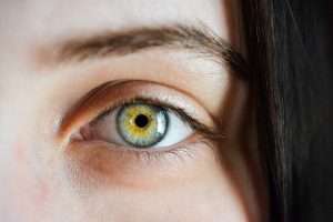
The eyes allow us to see and interpret the shapes, colours, and dimensions of an object. The eye can detect bright and dim light but is the most sensitive to visible light of wavelength 5500 Angestrum i.e., yellow light.
- Our eye is present in the eyeball.
- One eye covered a 150-degree angle.
Human Eye Diagram
Draw the labelled diagram of the human eye and explain the human eye structure:
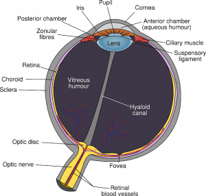
Main parts of the Human Eye
Cornea: A thin membrane covering the surface of the eyeball through which the light enters into the eye.
Iris: A dark muscular diaphragm located just behind the cornea.
Pupil: A black opening between the aqueous humour and eye lens. It regulates and controls the amount of light entering the eye.
- The hole in the iris is called the pupil.
- The pupil is also called a black spot.
- Light enters after the cornea.
- The size of the pupil is controlled by the iris.
- In sunlight, the size of the pupil is smaller.
- In the night, the size of the pupil is larger.
Ciliary muscles: Modify the curvature and focus of the eye lens. Hold the eye lens in position.
Crystalline lens: Composed of fibrous, jelly proteinaceous-like material covering in nature.
Retina: A real membrane having an enormous number of light-sensitive cells. It acts like a screen.
Sclera: The white part of the eye is called the sclera. The sclera is the outer layer of the eye.
Choroid: It provides the nutrition of our eyes. The choroid is made from blood, glucose and nutrition.
The function of the Human Eye
Light waves of an object enter the eye first through the cornea, a clear dome-shaped structure in the front of the eye. The cornea is responsible for the maximum refraction of light incidents on the eye. The light then passes through the pupil, the circular opening in the centre of the coloured iris.
Fluctuations in the intensity of incoming light change the size of the eye’s pupil. The pupil controls the amount of light that enters the eye. As the light entering the eye becomes brighter, the pupil constricts, due to papillary light response while as the light gets dimmer, the pupil dilates.
The crystalline biconvex lens that is located behind the iris and pupil, image is inverted and reserved. From here, the light continues through the vitreous humour, the clear gel that through the vitreous humour, the clear gel that makes up 80 per cent of the eye volume, and ideally, back to a clear focus on the retina.
The smallest area of the retina called the macula provides the best vision of any location in the retina. Within the layers of the retina, light impulses are changed by rod cells and cone cells into electrical signals to be carried by the optic nerve to the brain for interpretation.
Defects of the Human Eye
The human normally suffers from myopia, hypermetropia, astigmatism and old-age presbyopia.
| Myopia | Hypermetropia |
| 1. Near-sightedness, a person can clearly see nearby objects but not distant objects. 2. The image is formed in front of the retina. 3. The focal length of the eye lens increases. 4. It is corrected by the concave lens of suitable focal length/power. | 1. Far-sightedness, a person can clearly see distant objects but not nearby objects. 2. The image is formed behind the retina. 3. The focal length of the eye lens decreases. 4. It is corrected by the convex lens of suitable focal length/power. |
Astigmatism
The inability of the human eye to focus on objects in both horizontal and vertical lines is called astigmatism. This is caused due to varying curvatures in the eye lens in horizontal and vertical lines. In this defect, one or more surfaces of the cornea or eye lens are not spherical but, instead, are rough.
As a result of, there is no distant focus inside and the person fails to visualize the proper dimension of the objects. It is corrected by using cylindrical lenses.
Presbyopia
Among humans after the age of 45 years, eyes are most likely to be affected by Presbyopia because of the lessening flexibility of the eye lens along with weakening ciliary muscles which control the eye lens focal length. It is corrected using glasses having a bifocal lens in which the upper part is concave and the lower part is a convex lens.
Colour-Blindness
Cones with specific colours are there in the retina. If some cones are absent, the distinction of colours is not possible. In such a case, the person is said to be colour-blind. This defect arises due to the absence of colour responding to cone cells in the retina and due to genetic disorders. No cure has been developed till now by science.
Night-Blindness
Some persons have difficulty seeing objects in dim light during the night. This defect of the eye is called night blindness. This defect arises due to a lack of vitamin A and improper functioning of rod-shaped cells. The rod-shaped cells respond to intensity variation in light. So, by taking the proper amount of vitamin A in the diet, the functioning of rod-shaped cells may be improved.
Cataract
Sometimes, the crystalline lens of the eye, at old age, becomes milky land cloudy due to the growth of a thin membrane over it. This causes a partial or complete loss of vision. This condition is called a cataract. This is corrected by the surgical removal of extra growth.
Braille system for blind
It was developed by Louise Braille in 1821 but was adopted years later in 1932. The system has 63 dot patterns or characters in which each character represents a letter- these patterns are embossed on a Braille sheet to recognize them by touching with dots slightly raised, In India Ravindra Jain obtained his Sangeet Prabhakar degree, Lal Advani, himself visually challenged represented India in UNESCO on Braille problem. Helen A. Keller an American author and lecturer wrote the book Titled “Story of My Life”.
We hope guys, you enjoy this post about the human eye and the structure and function of the human eye. In the next post, discuss the working of the eye, and the construction of the eye.
Hello Sir, I am the student of class 10
Your post article is very helpful
Please update regular content and provide complete chapter of class 10
ok kamlesh visit regularly for more content for class 10
I like this article