The function of the human digestive system: The function of the digestive system is digestion and absorption of food. The mechanical and chemical breakdown of large, complex, non-diffusible food into small, simple, diffusible, and soluble forms is known as digestion.
Mechanical digestion: The breakdown of food with the help of digestive parts without chemical action is known as mechanical digestion.
Chemical digestion: The breakdown of food with the help of various enzymes that are present in the digestive tract is known as chemical digestion.
Example: Mixing food in the mouth by the tongue.
What is digestion?
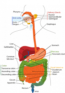
The breakdown of complex, water-insoluble food into small pieces, which are water-soluble and can be absorbed by the body, is called digestion. Human beings have an alimentary canal in which the food is digested and absorbed by the blood. This alimentary canal starts with the mouth and ends up in the anus. The main parts of the alimentary canal are the buccal cavity, esophagus, stomach, small intestine, large intestine, and anus. The liver and pancreas are other organs that contribute to the process of digestion.
Human digestive system
The organs which are responsible for ingestion, digestion, absorption, assimilation, and egestion constitute the digestive system. The digestive system comprises the alimentary canal and associated digestive glands. The alimentary canal in man is 9 meters long.
Diagram of the human respiratory system
Parts of the digestive system
The parts of the digestive system are:
- Buccal Cavity and Mouth
- Pharynx
- Oesophagus
- Stomach
- Small intestine
- Large intestine
Buccal Cavity and Mouth
The mouth also called the oral cavity or buccal cavity is the beginning of the alimentary canal. It is the upper part of where the digestion starts. Three main organs help in digestion in the buccal cavity. They are teeth, tongue, and salivary glands.
Teeth
The teeth are the hardest part of the body. They help in cutting, tearing, and grinding the food in the mouth. The well-ground food is easy to digest and move in the alimentary canal.
There is a set of 32 teeth in an adult mouth, i.e., 16 in each jaw (some may have 28 teeth as 4 wisdom teeth come late or may not come at all). Depending on their shapes and functions, teeth have been classified as:
Incisors (8 teeth): 4 + 4 teeth located in the middle of the upper and lower jaws. They are used for cutting the food.
Canines (4 teeth): 2 pointed teeth in each jaw separated by its incisors. They are used for the tearing of food.
Premolars (8 teeth): 4 teeth in each jaw, 2 on either side between the canines and molars. They are used for grinding the food.
Molars (8 teeth): 4 teeth in each jaw, 2 on either side. They are somewhat flat and are present at the rear of the mouth. Molars also grind the food.
Wisdom teeth (4 teeth): One each found at the end of each jaw. These teeth erupt at a later stage (at the age of 18+). Most of the time they don’t find enough space in the mouth and so are removed by the dentist. If present, they also help in grinding the food.
Children have a set of 20 temporary teeth or milk teeth. They don’t have molars. These teeth fall as children grow and are replaced by 32 permanent teeth.
Pats of Teeth
Enamel: The hardest bodily tissue covering the surface of the dental crown. It is as hard as crystal.
- Most shiny portion.
- Visible portion.
Dentine: Tissue that forms the tooth from the dental crown to the tooth root, situated inside the enamel and cementum is dentin. Dentine is softer than enamel.
Dental pulp: This tissue is called the nerves. Blood vessels and lymph vessels, as well as nerve fibers, are located in the dental pulp, supplying nutrients to the dentine. It connects with the brain.
Tongue
The tongue is a small muscular organ in the mouth that tells the taste of the food and helps to mix saliva with the food. It also helps to push the food into the esophagus through the pharynx.
- Length = 10 cm
- Mixing of saliva with food.
- The tongue is covered with moist, pink tissue called Mucosa.
- Tiny bumps called papillae give the tongue its rough texture.
- Thousands of taste buds cover the surface of the papillae.
- Taste buds are a collection of nerve-like cells that connect to the nerve running into the brain. Taste buds are more common in females.
The surface of the tongue is rough. Several taste buds on it help identify sweet, salty, sour, and bitter tastes. The taste buds for identifying different tastes are clustered in different regions of the tongue.
Lips
It doesn’t let liquid food and saliva come out of the mouth. The only organ that is not oily.
Oesophagus
It is a tube that connects the throat and the stomach. It helps in the transportation of food from the buccal cavity to the stomach. The length of the esophagus is 25 cm.
When the food is pushed from the mouth by the tongue into the pharynx, the food pipe opens. The pharynx opens into two pipes- the windpipe which is usually open and the food pipe or the esophagus which opens only when food comes. When food comes, epiglottis covers the windpipe and food enters the food pipe. From the esophagus, food moves down to the stomach by the contraction and expansion of the muscles of the food pipe. This type of movement is known as the peristaltic movement.
Stomach
The stomach secretes acids and enzymes that digest food. When food reaches the stomach, it is mixed with hydrochloric acid secreted in the stomach. This acid destroys the bacteria if present in the food and also makes the food acidic. It also secretes mucous which prevents its inner wall from the corrosive action of strong acid present inside. Digestive juice secreted by the stomach acts on the acidic food and breaks down proteins present in the food into simpler compounds.
- The shape of the stomach = J shapes
- Color of stomach = Red
- Three parts of the stomach are cardia, fundus, and pyloric.
- The gastric gland is present in the stomach.
- pH value of HCL is present in the stomach = 1.5 to 2.5.
The stomach secretes enzymes:
Pepsin: It breaks down the protein in food into small particles.
Renin: Converts milk into curd.
Mucin: It reduces the HCL effect. It protects the stomach lining from its acid.
Small intestine
The primary function of the small intestine is the absorption and complete digestion of nutrients and minerals from food using a small finger-life projection called villi. Villi provide surface area to the small intestine. Food moves onto the small intestine which is about 7.5 meters long.
Parts of Small Intestine
- Duodenum
- Jejunum
- Ileum
Duodenum
The first part of the small intestine is a c-shaped structure called the duodenum. In the duodenum, the liver pours its alkaline bile juice stored in the gall bladder through a bile duct. The acidic semi-fluid food (known as chyme) coming out of the stomach becomes alkaline when alkaline bile juice mixes with it in the duodenum. Bile juice emulsifies fats present in the food that is large globules of fats are converted to small globules for the better action of digestive enzymes.
There is another duct opening into the duodenum. It is the pancreatic duct that brings several enzymes from the pancreas and mixes them with chyme. The pancreatic juice contains enzymes like trypsin, lipase, amylase, chymotrypsin, etc. These enzymes can act only in an alkaline medium. Trypsin and chymotrypsin break peptides into amino acids, lipase breaks fats into fatty acids and glycerol, and amylase converts sugars into glucose. This process of digestion is done in the small intestine.
Jejunum
The digested food is observed by the walls of the small intestine, mainly the middle part which is the jejunum. The small intestine is specially designed for absorption. The inner wall of the small intestine contains many villi. The epithelial cells that line these villi have microvilli. These villi and microvilli help to increase the surface area of the intestine for the better absorption of food. There are several thin blood capillaries in the villi that bring the absorbed food into the blood. These capillaries join to form veins that carry blood towards the liver and from there to the heart.
From the heart, the digested food is transported by the blood to different organs of the body. In each cell of the body, the food is oxidized and energy is produced which helps us to do work.
Ileum
It is the lower part of the small intestine. The function of the ileum is to absorb vitamin B12 and bile salt and whatever products of digestion were not absorbed by the jejunum.
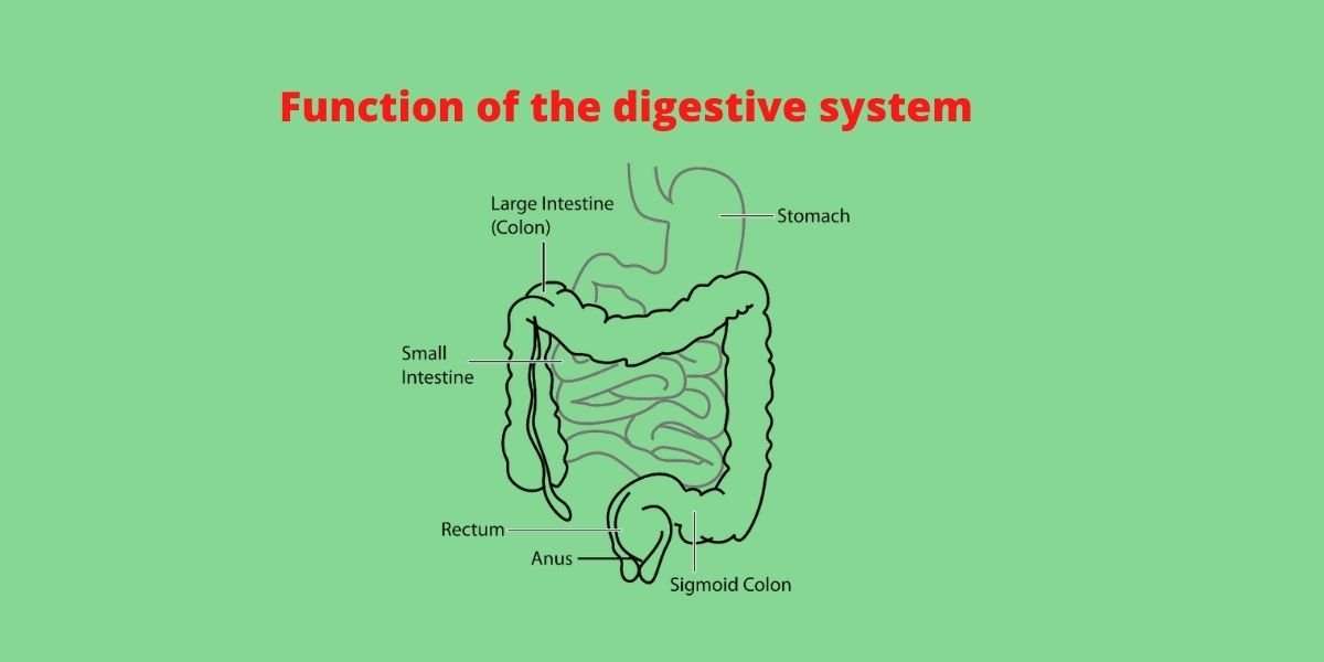
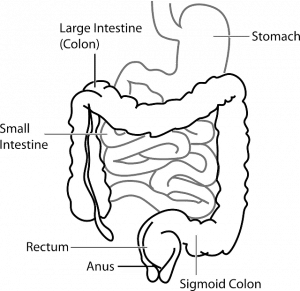
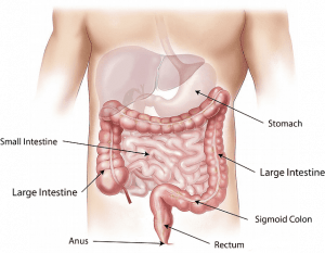
Large intestine
The undigested food is passed on to the large intestine. It is about 1.5 m in length. It represents the end of the digestive tract. Here excess water and some useful salts are absorbed. It reduces acidity, produces antibodies, and protects from infection. In the large intestine, the mucous membrane acts as a protective layer that prevents harmful bacteria from being reabsorbed into the body.
Parts of the large intestine
Cecum: The large intestine is joined to the end of the small intestine at the cecum.
Colon: It absorbs vitamin K that is created by colonic bacteria known as E.coli.
Rectum: It is the final segment of the large intestine that connects the colon to the anus.
Anus
The remaining waste in the form of semi-solid mass is stored in the rectum. From there, it is passed out through the anus. This process is called egestion.